Columbia Department of Radiation Oncology
Cutting-edge cancer care, education, and research in radiation treatment.

Radiation Oncology
Welcome to the Department of Radiation Oncology at Columbia University Irving Medical Center.
We combine cutting-edge radiation research and treatment with compassionate, personalized care. Our Department is home to the prestigious Center for Radiological Research (CRR), the nation's largest and most experienced university-based research center for radiation biology, where world-renowned doctors and scientists strive to move cancer treatment forward through focused research into the uses and biological effects of ionizing radiation.
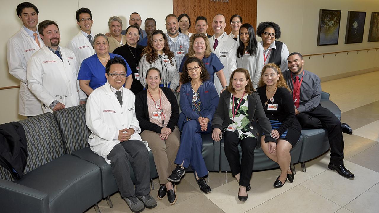
We collaborate with Columbia specialists in every field of medicine to provide world-class care at locations across the New York metro area.
Personalized Patient-Centered Care

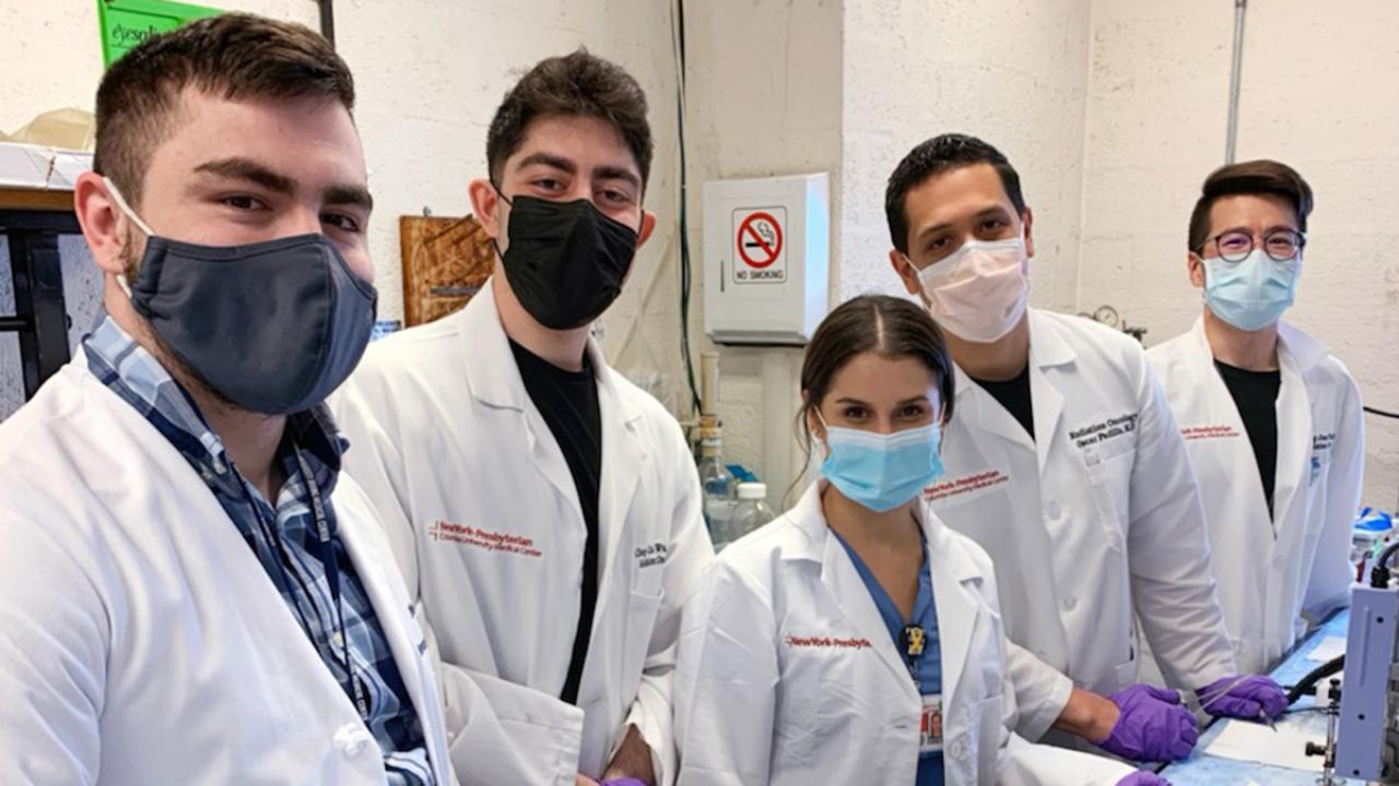
We tailor treatment to each individual using a highly coordinated multidisciplinary approach with leading experts, renowned surgeons, and the top medical oncologists.